Dr Ivan L. Simoneau
580 California St., Suite 400
San Francisco, CA, 94104
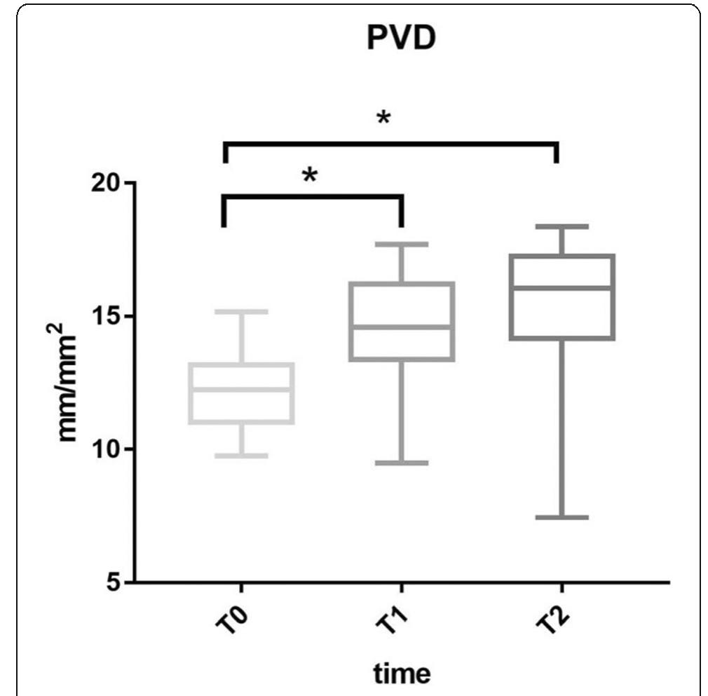
Introduction: Conventional assay technique to quantify vascular permeability in animal studies requires sacrifice animals; this becomes a barrier to evaluate of temporal changes or responses to therapeutic approaches in a single individual. In vivo fluorescence imaging potentially quantifies vascular permeability without sacrifice animals. However, the use of this noninvasive approach for the assessment of vascular permeability in remote organ injury caused by systemic inflammatory disease such as sepsis has not been reported. Methods: Cecal ligation and puncture (CLP)-induced septic mouse model was compared to sham and hydrocortisone pretreated (CLP + HC) mouse models. The lung was assumed as an injured remote organ and the footpad was assumed as a noninvasive observational site. The mixture of Evans blue (EB) and fluorescent dye of Genhance 750 were injected into mice, and the extraction of EB in harvested lung was assessed as a conventional indicator of vascular permeability. Fluorescent intensities in the harvested lung or footpad were assessed and their correlation was analyzed to investigate this novel, noninvasive approach to estimation of lung vascular permeability. Results: EB extraction in the harvested lung in the CLP group was significantly higher than in the other groups (CLP vs. sham, P=0.0012; CLP vs. CLP + HC, P=0.011). Fluorescent intensity in the footpad and harvested lung in the CLP group was also significantly higher than in the other groups (footpad, CLP vs. sham, P<0.0001; CLP vs. CLP + HC, P=0.0004; lung, CLP vs. sham, P<0.0001; CLP vs. CLP + HC, P<0.0001). The fluorescent intensity of the footpad was strongly correlated with that of the lung (r=0.95). Conclusions: The fluorescence imaging technique may be useful for assessment of vascular permeability based on EB quantification. The footpad fluorescent intensity was strongly correlated with that of the lung, and may be a suitable indicator in noninvasive estimation of lung vascular permeability.


![Conclusions: In clAls an appropriate empirical antibiotic therapy and an early infection source control are closely associated with better outcomes. score at infection which was higher in IAAT (p=0.04). Secondary peri- tonitis was the main type of clAl (45.5% in IIAT and 40.9% in IAAT) followed by abdominal abscess and biliary tract infection. Secondary bacteraemia was significantly higher in IIAT (p=0.03). Conversely, IAAT had an higher rate of adequate source control (p=0.01). Empi- rical therapy of IAAT patients included more frequently anti gram- positive (p=0.016) and carbapenems (p=0.01), while empirical dual anti gram negative and antifungal coverages rate were not different (Fig. 1). MDR and polimicrobial infections rate was significantly higher in II|AT when associated with septic shock at occurrence of infection (p=0.03; Fig. 2). IAAT showed significantly lower mortality at 28 and 90 days (p<0.01) as well as higher rate of clinical cure and microbio- logical eradication than IIAT (p<0.01). At the multivariate analysis, adequate source control [p=0.04, OR 0.25 (0.09-0.65)] and IIAT [(p<0.01, OR 11.4 (4.02-32.3)] turned out to be independently related with 28 days mortality. P065](https://figures.academia-assets.com/118529639/figure_029.jpg)


![Table 1 (abstract PO78). Bacterial count in lung, spleen and blood at euthanasia. Median [IQR]](https://figures.academia-assets.com/118529639/table_013.jpg)


![Fig. 1 (abstract P126). Ninety-day mortality among patients in the LEVO-CTS trial [3] in the subgroup of isolated CABG patients (n=563)](https://figures.academia-assets.com/118529639/figure_052.jpg)


![[1] Joosten et al. Anesthesiology 128:55-66, 2018](https://figures.academia-assets.com/118529639/figure_168.jpg)