Academia.edu no longer supports Internet Explorer.
To browse Academia.edu and the wider internet faster and more securely, please take a few seconds to upgrade your browser.
AI
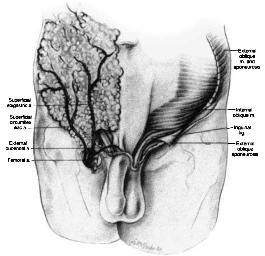
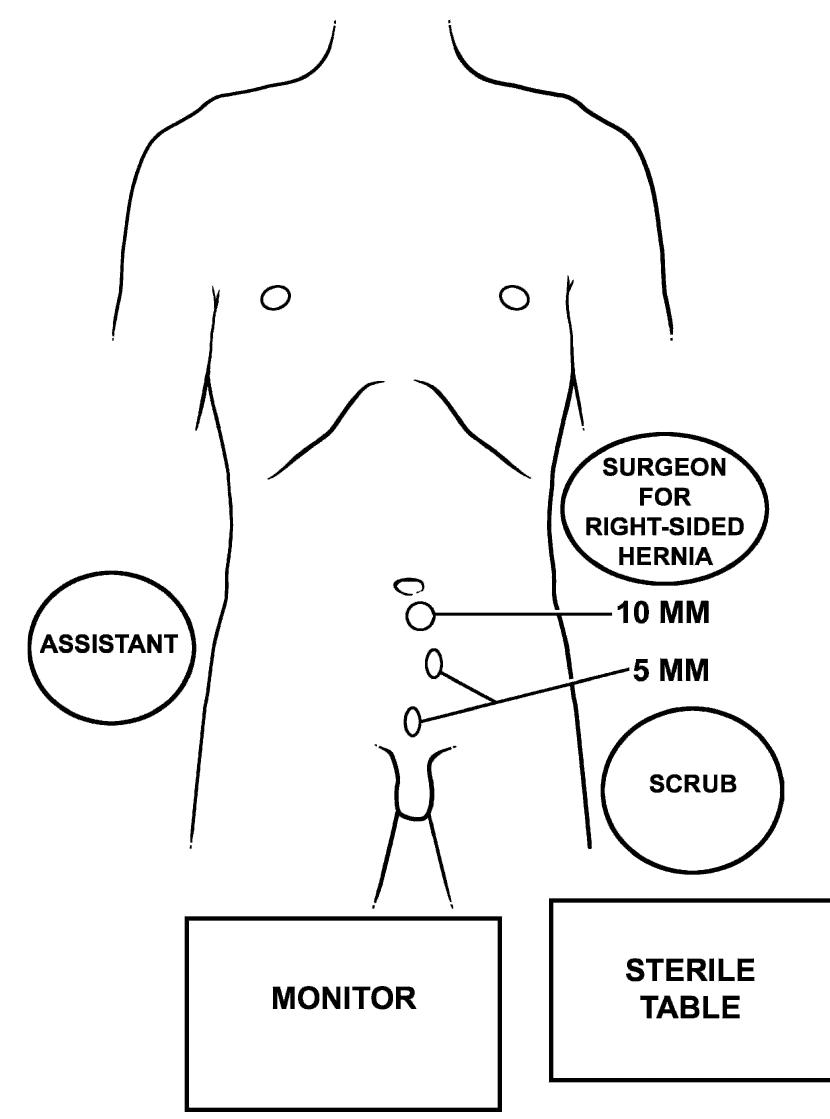
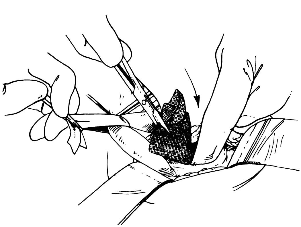
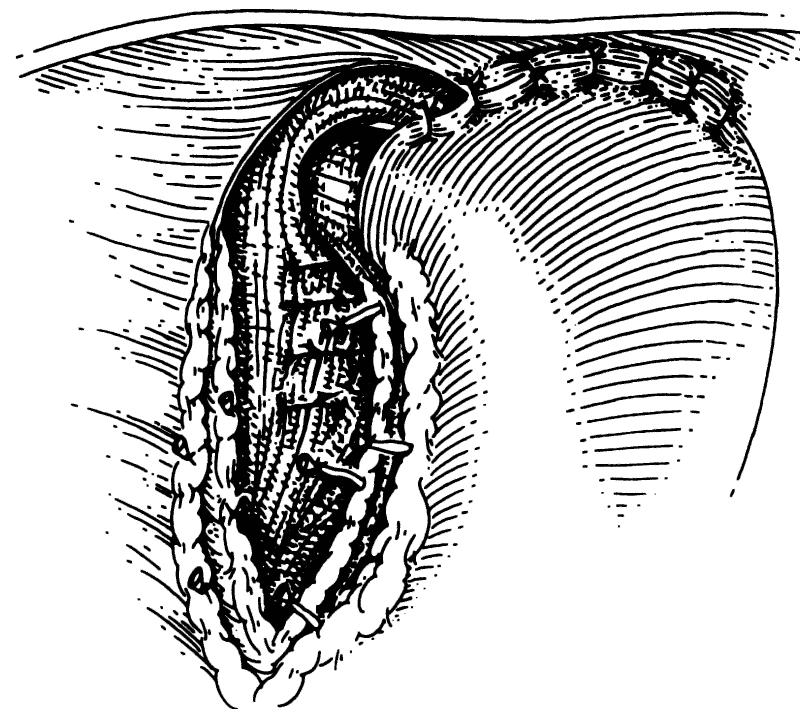
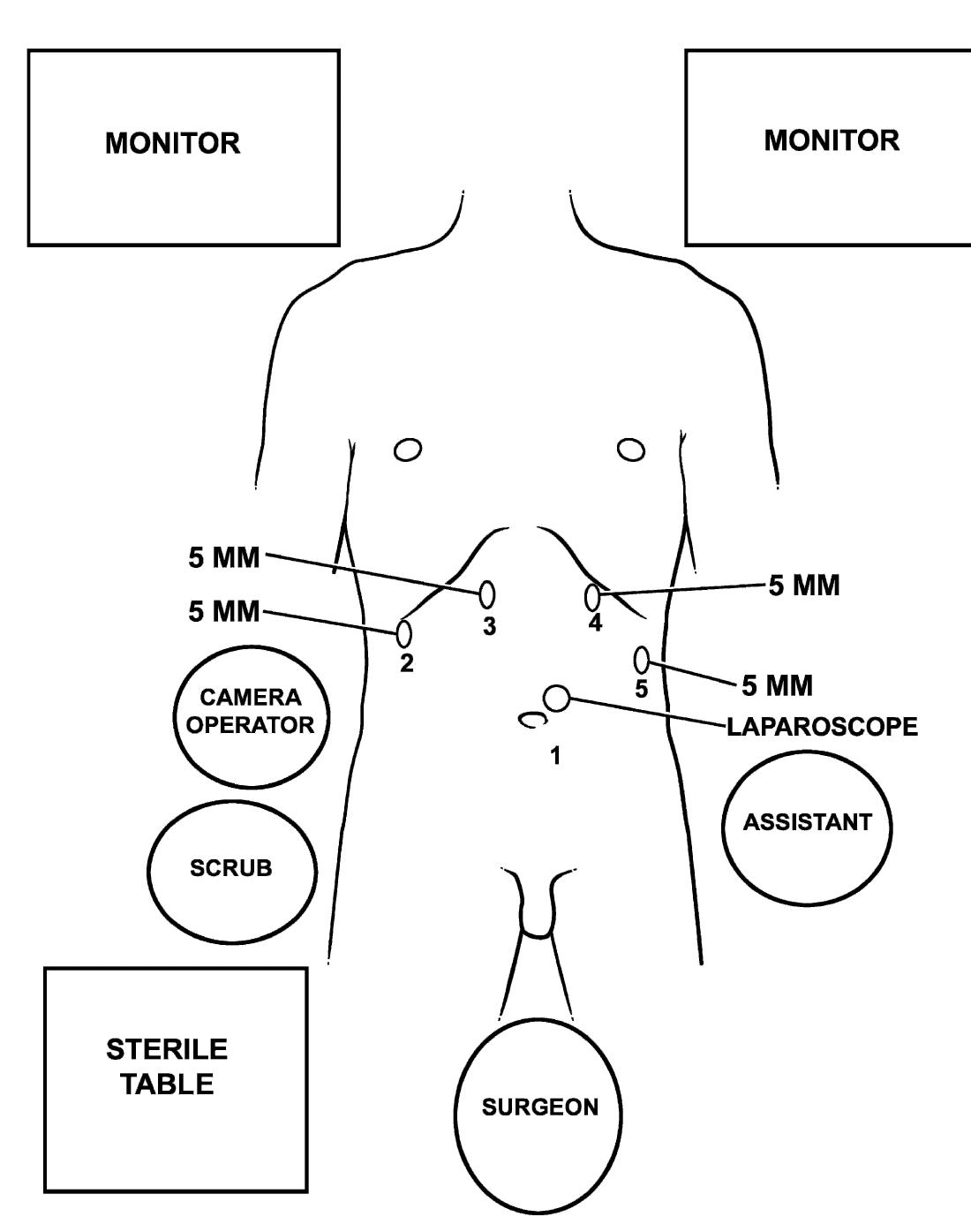
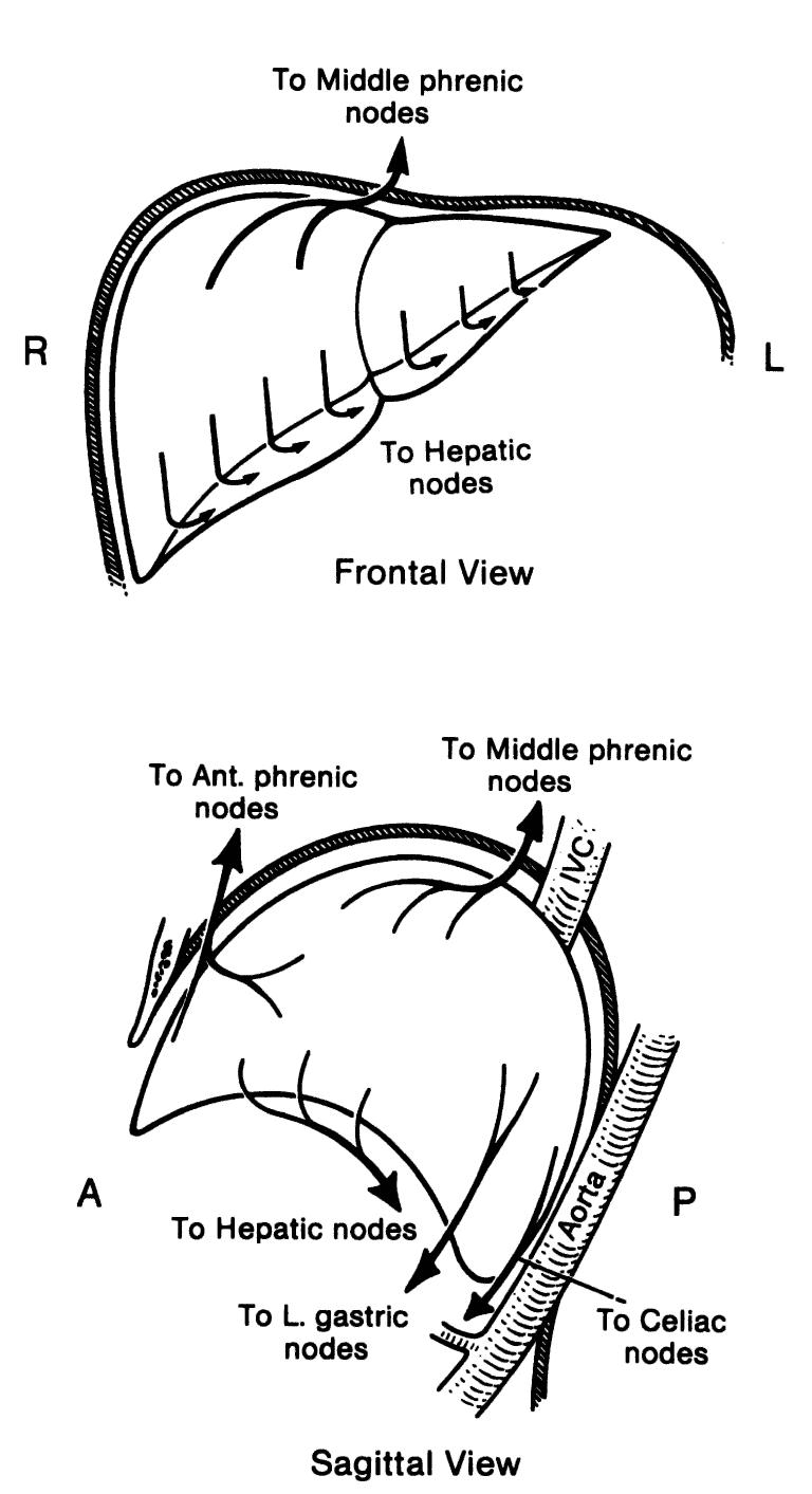
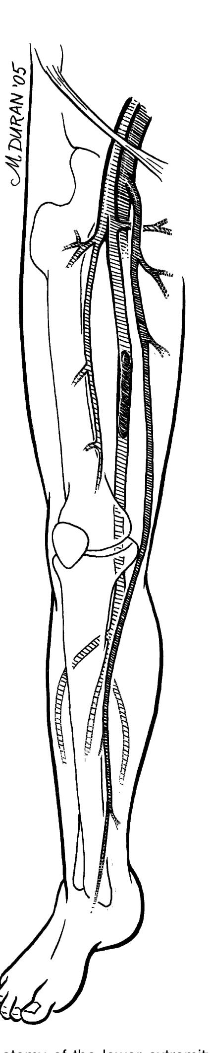
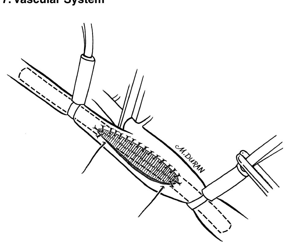
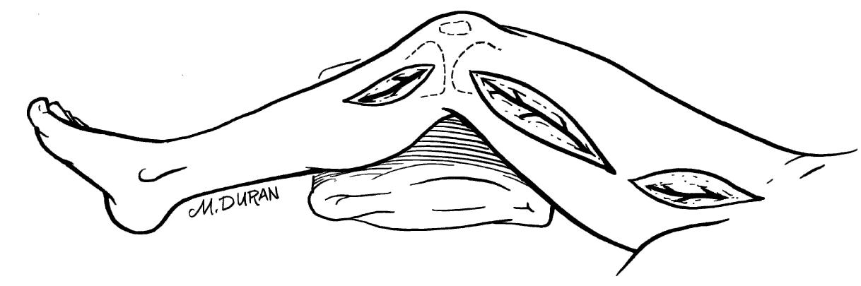
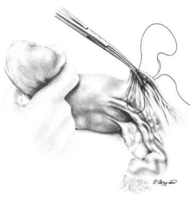
The paper discusses the surgical anatomy and techniques associated with various procedures, with particular emphasis on the anatomy of the scalp, including its vascular, lymphatic, and nerve supplies. It presents detailed insights into the blood supply of the scalp, highlighting the important arterial branches and their anastomoses, as well as the implications for surgical procedures. Additionally, the paper outlines techniques for surgical interventions, positioning, and considerations necessary for effective outcomes.
Plastic and Reconstructive Surgery, 1991
Journal of Plastic, Reconstructive & Aesthetic Surgery, 2012
Background: Total scalp avulsion is a rare and devastating event. Microsurgical replantation is the sole method to achieve an ideal cosmetic outcome. In the literature, most studies have reviewed limited sample sizes. Most authors report better outcomes when a greater number of microvascular anastomoses are used. This strategy remains controversial, as some authors have suggested that one artery may be sufficient. Methods: From 2005 to 2008, seven patients who sustained scalp avulsion underwent microsurgical replantation. All of the vascular anastomoses were made with a branch of the superficial temporal artery. We did not use vein grafts. The ischaemia time was 4e16 h. Results: In six cases, a single artery and one to two veins were anastomosed; in another case, two arteries and three veins were anastomosed. Six of the seven scalp replantations were successful and achieved normal hair regrowth. In five of the six successful cases, we performed a single-artery anastomosis. Conclusion: Scalp avulsion is rare around the world but occurs relatively frequently in our country due to the lack of safe and secure working conditions in agriculture and industry. In cases where multiple arterial anastomoses are not possible, the present findings suggest that one branch of the superficial temporal artery may be sufficient to reperfuse the replanted scalp and achieve excellent aesthetic results.
Injury, 1993
The freafmenf of choice for the avulsed scalp is microvascular replanfafion. Affer replanfafion there are numerous arterial anasfomoses, with venous congestion. UstMlly an effort to establish it&w through fwo arferial anasfomoses is made. We elected to anasfotnose one artery and mulfipk veins in each of our pafienfs. We present three consecufive successful replanfafions of total scalp au&on, using only one arterial anasfomosis, and mulfiple veins. Surprisingly, fwo of the patients regained their abilify to elevate fheir eyebrows actively.
Microsurgery, 1993
A successful replantation of a totally avulsed scalp. including both eyebrows, with only one arterial and one venous anastomosis to the superficial temporal vessels is described. Apart from a small partial skin necrosis of the right eyebrow, the entire transplant survived. Subsequently, the patient required only minor additional skin grafting, correction of scars hair micrografts. Replantation of the total scalp based on two or more vessels has previously been recommended and reported elsewhere. In the present case, complete survival of the scalp on only one artery and one vein was demonstrated, indicating that replantation should be considered even if available vessels for anastomosis are minimal. 8 1993 Wiley-uss. Inc. with a skin expander, and reconstruction of the eyebrow with MCROSUROERY 1 4-1SS3 Es wird die erfolgreiche Replantation cines komplett ausgerissenen Skalps einschlieSlich beider Augenbrauen iiber eine Anastomosierung der A. und V. superficialis temporalis vorgestellt. AuOer einer kleinen Teibkrose der Haut im Bereich der rechten Augenbraue blieb das gcaamte Transplantat vital. Im weiteren Verlauf m t i g t e die Patientin lediglich geringfiigige ZuSiItzliche Hauaransplante, eine Narbenkorrektur mittels Hautexpander und die Rekonstruktion einer Augenbraue mit Minihaamansp lantaten. Die Replantation des gesamten Skalps iiber zwei oder meh GeFaSe wurde bereits friiher empfohlen und auch an anderer Stelle berichtet. Im vorliegenden Fall wurde gezeigt, das der Skalp in toto vital blieb bei nur einer Anastomose einer einzigen Arterie und einer einzigen vene. somit konnte ndchgewiesen werden, das eine Replantation unbedingt ins Auge gefaRt werden SOU, selbst wenn nur spiirliche Gefiiiiverhiiltnisse zur Anastomosierung vorhanden sind.
Plastic Surgery, 2010
Surgical and …, 1992
The arrangement and structure of the fascial layers of the scalp were studied in 48 red neoprene latex-injected cadavers with the aid of an operating microscope. The galea aponeurotica was continuous with a superficial temporal fascia. Deep to the galea, the subaponeurotic connective tissue was bilaminar. There was an outer, vascular, areolar layer, and an inner, avascular, membranous layer. Underlying the temporalis muscle, the pericranium was thinner and more adherent than elsewhere with no subpericranial tissue.
European Journal of Plastic Surgery, 1997
The authors report their experience in successful reimplantation of avulsed scalp in two patients with one arterial and one venous anastomosis to the superficial temporal vessels. In both cases a double vein graft, harvested from the dorsum of the hand, was interposed between the pedicle of the scalp and the recipient temporal vessels to avoid tension after trimming of the damaged segments. Scalp replantation based on two or more vessels has previously been reported in the literature. In the present cases, the complete survival of the scalp on only a single vascular pedicle suggests that replantation should be considered also when the available vessels for the anastomosis are few. Moreover, even if these reconstructions are lengthy, a prolonged ischemic interval does not appear to be a significantly limiting factor for the success of the revascularization. The cosmetic and psychological success of these cases lend support to the idea that one should always attempt a microvascular replantation of avulsed scalps.
Clinical Anatomy, 2010
Microsurgery, 2012
Background: Soft tissue defects of the scalp may result from multiple etiologies and can be challenging to reconstruct. We discuss our experience with scalp replantation and secondary microvascular reconstruction over 36 years, including techniques pioneered at our institution with twin-twin scalp allotransplant and innervated partial superior latissimus dorsi (LD) for scalp/frontalis loss. Methods: A retrospective review of all patients presenting with scalp loss requiring microvascular reconstruction at a single center was performed from January 1971 to January 2007. Medical records were reviewed for age, gender, defect size/location, etiology, type of reconstruction, recipient vessels used, vein grafts, and complications. Results: Thirty-three patients were identified; mean age was 33 years (range, 7-79). Mean scalp defect size was 442 cm 2 (range, 120-900 cm 2). Thirty-six microvascular reconstructions were performed; of these, 10 scalp replants and 26 microvascular tissue transfers. Of these 26, 17 were LD based (partial superior LD with and without reinnervation, LD combined with serratus, LD combined with parascapular, LD combined with split rib, LD only) and 2 free scalp allotransplant among others. The superficial temporal artery and vein was used as recipient vessels in 70% of cases. Overall, microvascular success rate was 92%; complications occurred in 14 cases, nine major (tumor recurrence [n 5 2], partial flap loss [n 5 2], replant loss [n 5 3, size <300 cm 2 ], hematoma [n 5 2]) and five minor (donor site seroma /hematoma [n 5 3], flap congestion [n 5 1], superficial wound infection [n 5 1]). Conclusions: Every attempt should be made at scalp replantation when the patient is stable and the parts salvageable. Larger avulsion defects had higher success rates after replantation than smaller defects (<300 cm 2), with the superficial temporal artery and vein most commonly used for recipient vessels (P 5 0.0083). Microvascular tissue transfer remains a mainstay of treatment for scalp defects, with LD-based flaps, demonstrating excellent versatility for a range of defects. V V
2019
The facial artery (a branch of the external carotid artery) is the main artery of the face. It gives rise to seven branches viz. inferior labial, superior labial, inferior alar, superior alar, lateral nasal and angular arteries, which are variable. This study included a dissection of twenty embalmed adult cadaveric head and neck specimens. The parameters of origin, branching patterns, termination and variations were analysed and compared with sex and laterality. The facial artery followed the standard anatomical description of origin in 84.62 % of the sample. Variations: (i) origin as a linguofacial trunk in 12.82 % and (ii) high origin in 2.56 % was observed. Male specimens displayed a higher number of linguofacial trunk origins (7.69 %). The branching patterns of the facial artery was classified into six types, with subtypes for Types 1 and 2. Subtype 1-A (standard anatomical description with early termination) occurred in most of the sample (46.15 %). Males were found to have more variations in branching patterns than females (48.72 % and 41.03 % respectively). Termination of the facial artery was as follows: inferior labial artery (5.13 %), superior labial artery (10.26 %), inferior alar artery (10.26 %), superior alar artery (46.15 %), lateral nasal artery (5.13 %), and angular artery (20.51 %). A single case (2.56 %) of an abortive artery was noted. Statistical analysis showed that sex was independent of each parameter observed in this study. Anatomical knowledge of the facial artery is of importance to clinicians and surgeons during procedures such as musculomucosal, island flaps and aesthetic dermatology.
Anatomical knowledge of the facial vasculature is crucial for successful plastic, reconstructive, and minimally invasive procedures of the face. The facial artery is palpated as it crosses the inferior mandibular rim immediately anterior to the insertion of the masseter muscle. This point is also helpful in locating the course of the mandibular branch of the facial nerve. After crossing the mandibular rim, the facial artery and vein then follow an anterosuperior course in the direction of the oral commissure. Near the angle of the mouth, the inferior labial artery and then the superior labial artery branch off medially The facial vein, also referred to as the anterior facial vein, is a paired vessel and the main vein of the face. The facial veins stem from the angular veins on each side of the root of the nose. Some sources claim that the angular vein, which arises from the confluence of the supratrochlear and supraorbital veins, is only an initial segment of the facial vein.
Annals of Plastic Surgery, 2011
The main arterial supply of the facial skin envelope is the facial artery which serves as the main pedicle for a number of facial flaps, including a facial transplant graft. This study explored the course of the facial artery and vein, branching patterns, terminations, and anomalous variants. Cadaveric dissections of 201 facial arteries and 198 facial veins were performed. All branches originated from a single facial arterial trunk in 86% of specimens and branching patterns were symmetrical in 53%. The facial artery predominantly terminated as a lateral nasal artery (49%). In 5 cases, the facial artery was undetectable with transverse facial arterial dominance (1 case bilateral). The facial vein was predictable in position except for 2 instances, being replaced by a transverse facial vein (unilateral). Facial arterial dominance in facial blood supply is common but unpredictable. Careful vascular workup prior to facial transplantation and unipedicled flap procedures is therefore essential.
Journal of Craniofacial Surgery, 2019
Complex scalp defects with poor recipient vessels represent a challenge for plastic surgeons. In these cases, free flaps are the last resort for solving the problem. The authors present
Surgery Today, 1994
A simple technique for the excision of vascular malformations of the scalp is described herein. Surgical excision is easily accomplished by placing hemostatic sutures all around the lesion outside the line of excision, whereby minimal blood loss is achieved. The resulting defect can be covered by a split skin graft or flap.
Th e aim of the study was to evaluate the terminal segmentation of the axillary artery and to present four cases of anomalous branching of the axillary artery, the superfi cial brachial artery (arteria brachialis superfi cialis), which is defi ned as the brachial artery that runs superfi cially to the median nerve. Totally, cadaveric upper arms embalmed by classical formaldehyde technique from collections of the Department of Anatomy, Th ird Faculty of Medicine, Charles University in Prague, were macroscopically dissected with special focus on the branching arrangement of the axillary artery. Th e most distal part of the axillary artery (infrapectoral part) terminated in four cases as a bifurcation into two terminal branches: the superfi cial brachial artery and profunda brachii artery, denominated according to their relation to the median nerve. Th e profunda brachii artery primarily gave rise to the main branches of the infrapectoral part of the axillary artery. Th e superfi cial brachial artery descended to the cubital fossa where it assumed the usual course of the brachial artery in two cases and in the other two cases its branches (the radial and ulnar arteries) passed superfi cially to the fl exors. Th e incidence of the superfi cial brachial artery in our study was of cases. Th e reported incidence is a bit contradictory, from . to of cases. Th e anatomical knowledge of the axillary region is of crucial importance for neurosurgeons and specialists using the radiodiagnostic techniques, particularly in cases involving traumatic injuries. Th e improved knowledge would allow more accurate diagnostic interpretations and surgical treatment.
Plastic and Reconstructive Surgery, 2009
Background: Nasal reconstruction with use of the forehead flap has been performed for hundreds of years. Forehead vasculature has been studied; however, anatomical relationships to the forehead flap have not been adequately examined. This anatomical study evaluated the vascular anatomy of the paramedian forehead flap. Methods: Five fresh cadaver heads were used. Four underwent cannulation of internal and external carotids bilaterally followed by injection of a barium sulfate/gelatin mixture and three-dimensional computed tomographic angiography to evaluate vascular anatomy. In one specimen, the supraorbital, supratrochlear, and angular arteries were cannulated. Methylene blue dye was injected to identify vascular territory followed by injection of contrast media for dynamic four-dimensional computed tomographic angiography. A paramedian forehead flap was raised and the injections were repeated. Colored-latex was injected followed by dissection. Measurements were made on a computed tomography workstation. Results: A periorbital plexus extends to 7 mm over the orbital rim. The angular, supratrochlear, and supraorbital arteries communicated into the flap by means of the vascular plexus. The supratrochlear vessel ran axially into the forehead flap and continued across the transverse limb of the flap. The deep branch of the supratrochlear ascended the periosteum under the flap. Noncontiguous vessels were noted to back-fill with latex through the subdermal plexus in the distal flap. Conclusions: Maximal three-vessel flow may be obtained by preserving periosteum at least 3 cm over the orbital rim and beginning the flap 7 mm above the orbital rim. The subdermal plexus of the forehead is robust, enabling preservation of the distal transverse limb of the forehead flap.
Cureus, 2020
The supratrochlear artery represents a terminal branch of the ophthalmic artery. Cosmetic interventions may traumatize it, resulting in a circulation in the lesion in glabellar region and in the medial aspect of the forehead. This review article aims to synopsise the existing knowledge of the anatomy of the supratrochlear artery in close correlation with minimally invasive cosmetic procedures in the facial area such as soft-tissue filler injections. Their possible adverse effects and their safe application based on the topographic anatomy were included. A literature review was performed in PubMed/Medline online medical database. The superficial course of the supratrochlear artery, as well as the rich, variable anastomotic network that it forms with the supraorbital, angular and dorsal nasal artery raise clinical questions in the case of soft-tissue filler injections in the nasoglabellar and central forehead area. Accidental cannulation of the supratrochlear artery and ultimately, the risk of embolization of the central retinal artery in a retrograde fashion might lead to injury with questionable cosmetic results. Although the risk of complications from the use of soft tissue fillers is considered rare, once happen, the results could be devastating for the quality of life. Thus, the comprehension of the anatomy of the supratrochlear artery is paramount for the health practitioners.
Current Concepts in Plastic Surgery, 2012
Bangladesh Journal of Anatomy, 2014
Context: Basilar artery is formed by the union of right & left vertebral arteries at the lower border of pons / mid medullary level which courses upward along Basilar groove on the anterior surface of pons & supplies the anatomical structures within the posterior cranial fossa of brain. The Circle of Willis is the principal arterial anastomotic trunk of brain formed by anastomosis between the branches of internal carotid arteries and terminal branch of basilar artery. It is responsible for collateral circulation of brain, specially of old people - who have reduced blood supply to brain due to senile arteriosclerosis. Cerebrovascular diseases due to basilar artery is more common in men than women. Anatomy of basilar artery and its branches are very complex and variable. So, detailed morphogical knowledge is essential for proper diagnosis and treatment of neurological disorder. To identify whether there is any variations in the location of basilar artery in relation to basilar groov...
Morphologie, 2019
Étude anatomique des territoires artériels de la face dépendants des branches de l'artère carotide externe
Loading Preview
Sorry, preview is currently unavailable. You can download the paper by clicking the button above.